 | Info
Sheets |
| | | | | | | | | | | | | | | | | | | | | | | | |
 | Out-
side |
| | | | |
|
| | | | |
Result : Searchterm 'spiral imaging' found in 0 term [ ] and 2 definitions [ ] and 2 definitions [ ], (+ 11 Boolean[ ], (+ 11 Boolean[ ] results ] results
| | previous 6 - 10 (of 13) nextResult Pages : [1] [2 3] |  | |  | Searchterm 'spiral imaging' was also found in the following service: | | | | |
| |  |
| |
|
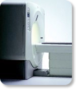
From GE Healthcare;
GE's Signa Contour/i system uses the innovations like K4 technology and real-time interactive imaging.
This compact magnet with wide-flare gantry obtains high patient comfort with low costs.
Device Information and Specification CLINICAL APPLICATION Whole body Head and body coil standard; all other coils optional; open architecture makes system compatible with a wide selection of coils Standard: SE, IR, 2D/3D GRE and SPGR, Angiography;; 2D/3D TOF, 2D/3D Phase Contrast;; 2D/3D FSE, 2D/3D FGRE and FSPGR, SSFP, FLAIR, optional: EPI, 2D/3D Fiesta, FGRET, Spiral2D 0.8 mm to 20 mm; 3D 0.1 mm to 5 mm 128x512 steps 32 phase encode POWER REQUIREMENTS 480 or 380/415 V STRENGTH SmartSpeed 23 mT/m, HiSpeed Plus 33 mT/m | | | | | |
| | | | | |
| |
|
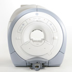
From GE Healthcare;
The GE Signa HDx MRI system is a whole body magnetic resonance scanner designed to support high resolution, high signal to noise ratio, and short scan times.
The 1.5T Signa HDx MR Systems is a modification of the currently marketed GE 1.5T machines, with the main difference being the change to the receive chain architecture that includes a thirty two independent receive channels, and allows for future expansion in 16 channel increments. The overall system has been improved with a simplified user interface
and a single 23" liquid crystal display, improved multi channel surface coil connectivity, and an improved image reconstruction architecture known as the Volume Recon Engine (VRE).
Device Information and Specification CLINICAL APPLICATION Whole body CONFIGURATION Compact short bore Standard: SE, IR, 2D/3D GRE and SPGR, Angiography: 2D/3D TOF, 2D/3D Phase Contrast; 2D/3D FSE, 2D/3D FGRE and FSPGR, SSFP, FLAIR, EPI, optional: 2D/3D Fiesta, FGRET, Spiral, Tensor, 2D 0.7 mm to 20 mm; 3D 0.1 mm to 5 mm 128x512 steps 32 phase encode POWER REQUIREMENTS 480 or 380/415 less than 0.03 L/hr liquid helium | | | | | |
| | | | | |
| |
|
Device Information and Specification CLINICAL APPLICATION Whole body Head and body coil standard; all other coils optional; open architecture makes system compatible with a wide selection of coils Standard: SE, IR, 2D/3D GRE and SPGR, Angiography;; 2D/3D TOF, 2D/3D Phase Contrast;; 2D/3D FSE, 2D/3D FGRE and FSPGR, SSFP, FLAIR, optional: EPI, 2D/3D Fiesta, FGRET, SpiralTR 4.4 msec to 12000 msec in increments of 1 msec TE 1.0 to 2000 msec; increments of 1 msec Simultaneous scan and reconstruction;; up to 100 images/second with Reflex 100 2D 0.7 mm to 20 mm; 3D 0.1 mm to 5 mm 128x512 steps 32 phase encode 0.08 mm; 0.02 mm optional POWER REQUIREMENTS 480 or 380/415 V Less than 0.03 L/hr liquid heliumSTRENGTH SmartSpeed 23 mT/m, HiSpeed Plus 33 mT/m 4.0 m x 2.8 m axial x radial | | | |
• View the DATABASE results for 'Signa Infinity 1.0T™' (2).
| | | | |
| | | Searchterm 'spiral imaging' was also found in the following service: | | | | |
| | |
| |
|

From GE Healthcare;
three dedicated MRI systems - a neuro imager, a cardiovascular system, and a whole body scanner - all in one system.
Device Information and Specification CLINICAL APPLICATION Whole body Head and body coil standard; all other coils optional; open architecture makes system compatible with a wide selection of coils Optional 2D/3D brain and prostate Standard: SE, IR, 2D/3D GRE and SPGR, Angiography: 2D/3D TOF, 2D/3D Phase Contrast;; 2D/3D FSE, 2D/3D FGRE and FSPGR, SSFP, FLAIR, EPI, optional: 2D/3D Fiesta, FGRET, Spiral, TensorTR 1.2 to 12000 msec in increments of 1 msec TE 0.4 to 2000 msec in increments of 1 msec 2D 0.7 mm to 20 mm; 3D 0.1 mm to 5 mm 128x512 steps 32 phase encode POWER REQUIREMENTS 480 or 380/415 Less than 0.03 L/hr liquid He STRENGTH Zoom 40 mT/m, whole body 23 mT/m 4.0 m x 2.8 m axial x radial | | | |
• View the DATABASE results for 'Signa Infinity 1.5T™ TwinSpeed with Excite' (2).
| | | | |  Further Reading: Further Reading: | News & More:
|
|
| |
| | | | | |
| |
|
Flow phenomena are intrinsic processes in the human body. Organs like the heart, the brain or the kidneys need large amounts of blood and the blood flow varies depending on their degree of activity. Magnetic resonance imaging has a high sensitivity to flow and offers accurate, reproducible, and noninvasive methods for the quantification of flow. MRI flow measurements yield information of blood supply of of various vessels and tissues as well as cerebro spinal fluid movement.
Flow can be measured and visualized with different pulse sequences (e.g. phase contrast sequence, cine sequence, time of flight angiography) or contrast enhanced MRI methods (e.g. perfusion imaging, arterial spin labeling).
The blood volume per time (flow) is measured in: cm3/s or ml/min. The blood flow-velocity decreases gradually dependent on the vessel diameter, from approximately 50 cm per second in arteries with a diameter of around 6 mm like the carotids, to 0.3 cm per second in the small arterioles.
Different flow types in human body:
•
Behaves like stationary tissue, the signal intensity depends on T1, T2 and PD = Stagnant flow
•
Flow with consistent velocities across a vessel = Laminar flow
•
Laminar flow passes through a stricture or stenosis (in the center fast flow, near the walls the flow spirals) = Vortex flow
•
Flow at different velocities that fluctuates = Turbulent flow
See also Flow Effects, Flow Artifact, Flow Quantification, Flow Related Enhancement, Flow Encoding, Flow Void, Cerebro Spinal Fluid Pulsation Artifact, Cardiovascular Imaging and Cardiac MRI. | | | | | |
• View the DATABASE results for 'Flow' (113).
| | |
• View the NEWS results for 'Flow' (7).
| | | | | | Further Reading: | News & More:
|
|
| |
| | | | |
| | |
| | | |
|
| |
 | Look
Ups |
| |


